Imaging
Receive the latest news, event invites, funding opportunities and more from the Ontario Institute for Cancer Research.
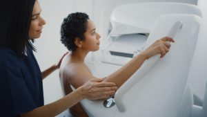
Imaging lets us see a patient’s cancer inside the body and gives us clues on where and how to treat it. The Imaging Program helps bring cancer imaging discoveries to doctors who can use them in hospitals and clinics.
There are many new imaging systems being developed in Ontario research labs. We want to move them into clinical use when ready because they have great potential to improve how cancer is diagnosed and treated.
We have four teams of experts moving imaging discoveries from the lab to the clinic as quickly as possible.
The Medical Imaging Instrumentation and Software team focuses on new ways to detect cancer early and tools to guide cancer treatment. These can help avoid common side effects like damage to nearby normal tissue. Many of the new tools can be used at the time and place where the patient receives care in a hospital.
The Imaging Validation Core team creates tools to measure and analyze cancer and nearby healthy tissue. Its researchers have developed unique approaches to study large tissue samples more efficiently. They look at the tell-tale features of cancer cells and nearby normal cells to identify what type and stage of cancer a patient has. This helps doctors customize a patient’s personalized treatment.
The Probe Development group makes molecules that are tagged with small amounts of radioactivity. When injected into the body, these molecules attach to cancer cells and make it possible to see their exact location in medical images. This team works with doctors and industry partners to bring their discoveries and products to cancer centres in Ontario and around the world.
The Quantitative Imaging for Personalized Cancer Medicine team helps to make it easier to use imaging in clinical trials. They help set standards for image quality and provide specialized software for image analysis. The team has also developed online software for sharing images across hospitals.
The mission of the Imaging Program is to build the capability in Ontario to accelerate translation of research findings and developments of new imaging techniques and probes of cancer into clinical practice through the critical stages.
We have 4 centres of excellence to provide the necessary expertise to the cancer research and clinical communities.
Please visit OICR’s Collaborative Research Resources directory for more opportunities to collaborate.
What we do
The Centre for Probe Development and Commercialization (CPDC) helps create new nuclear medical technologies that can help cancer patients live longer, healthier lives. We help universities and companies take ideas and inventions from initial concept to final government approval. We provide clinicians with new technologies that can be used for safer imaging and treatment options for cancer.
Why we do it
Nuclear medical technologies are ideal for finding cancer at early stages. Early detection gives a patient the best chance of starting a successful treatment plan. Once a tumour is found, nuclear technology can be used to focus the treatment at its exact location. This reduces side effects that are common with other unfocused treatments like older chemotherapies.
How we do it
Our focus is on using radioactive probes that are injected into patients. The radioactive probe will attach to the cancer, and light up in images so doctors can see the tumour. By knowing the size and location of the tumour, doctors can design the best treatment plan. Doctors can also safely watch how the cancer is reacting to the chosen treatment. We can also attach more powerful radioactive material to create probes for cancer treatment. These stronger probes will destroy the cancer cells when they bind to them.
More about Probe Development
Probe Development focuses on the discovery, development, distribution, and commercialization of molecular imaging probes and targeted radiotherapeutics. This work is carried out at the Centre for Probe Development and Commercialization (CPDC) locations in Hamilton, Toronto and Ottawa.
Expertise and capabilities
The Probe Development team has the expertise to overcome the gap between probe research and clinical use. The CPDC provides the cancer research community with the collaborative support they need to access existing and novel probes. Lay expertise includes radiopharmaceutical research and development, process development and manufacturing, quality assurance, regulatory affairs and business development.
Principal Investigator
Dr. John Valliant, Founder, CPDC; Professor, Department of Chemistry and Chemical Biology, McMaster University
Contact
Dr. Anne Goodbody,
Chief Regulatory Officer
Website
http://www.imagingprobes.ca/
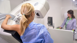
What we do
We work with researchers, doctors and companies to create new ultrasound imaging tools to detect, diagnose and treat cancer.
Why we do it
As people get older, most types of cancers become more common. This puts more pressure on our healthcare system. Finding cancer earlier can help ease this pressure. So it’s important to create better, less expensive ways to find cancer early that can be used when and where a patient receives care in a hospital.
The technologies we are developing have the potential to detect and treat cancer using an ultrasound machine instead of more expensive systems. This makes them better solutions for smaller and remote communities. They could also help decrease the number of people waiting for other types of scans to diagnose their cancers.
How we do it
We have invented robotics and machine learning approaches to diagnose, locate and treat tumours. These include:
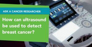
3D ultrasound diagnostic systems. We are developing new systems to diagnose cancer at the point-of-care by detecting cancer and quickly analyzing images of the disease. One device can detect cancer in women with dense breasts. Another can detect thyroid cancer by automatically analyzing images. A third can detect and assess oral cancer.
3D ultrasound-guidance systems. We are creating new ultrasound imaging tools to treat many cancers. In female reproductive system cancers, our system guides the placement of radioactive seeds so they can destroy tumour cells. A similar approach is possible for liver tumour cells. For prostate cancer, our new imaging method will help doctors make sure that the tools they use are aimed at the right place to treat the entire tumour while avoiding nearby healthy tissue.
More about Medical Imaging Instrumentation and Software
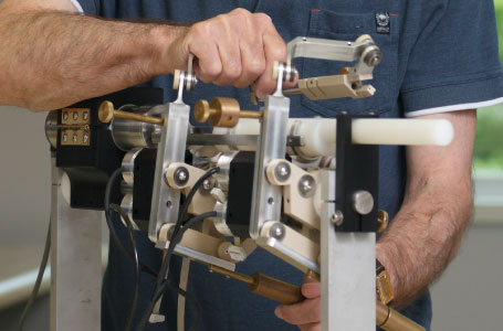
Medical Imaging Instrumentation and Software (MIIS) develops image-guided interventional imaging systems that are used in a wide range of applications, from multi-modality biopsy systems to image-guided robotic techniques used for minimally invasive focal tumour ablation. This work is carried out at the Centre for Imaging Technology Commercialization (CIMTEC) and the Robarts Research Institute.
Expertise and capabilities: The MIIS team has core expertise in imaging physics and mechanical, electrical and computer engineering, needed to develop image-processing, guidance, visualization tools and robotic devices that can be used in a wide range of cancer biopsy and therapy applications. We are have core technology for robotic devices, image guidance and processing tools that can be applied to emerging clinical needs.
Capabilities include 3D multi-modality image viewing, delineation of organ and tumour margins (segmentation), fusion of multi-modality images (registration), texture analysis of images (radiomics), 3D ultrasound (3D US) biopsy guidance instrumentation, robotic systems for image-guided therapy, and image-guidance software. Access to imaging equipment includes MR scanners, ultrasound machines and CT scanners as well as fully equipped machine and electronics shops.
Principal Investigator
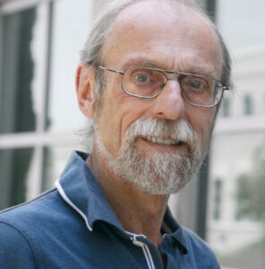
Dr. Aaron Fenster, CEO, CIMTEC; Chair, Division of Imaging Sciences, Department of Medical Imaging, Western University; Scientist, Robarts Research Institute; Professor, Departments of Biomedical Engineering, Oncology, Medical Imaging and Medical Biophysics, Western University;
Contact
Carol Richardson
Website
http://www.cimtecimaging.com/
What we do
The Imaging Validation Core (IVC) helps get new and better medical imaging systems and analysis methods into the clinic as quickly as possible. To do this, our lab uses images to perform in-depth studies of cancer cells and the tissue around them. We develop new ways to image cancer and improve analysis of these images. And we work with researchers who develop new tools for imaging to test if these tools can improve how cancers are found inside the human body. On a “big picture” scale, IVC is working to learn where a cancer starts, how it evolves, how aggressively it might grow and what is the most effective way to treat it.
Why we do it
Every person’s cancer is different, even if they have the same type of cancer, like breast cancer. Our work provides insight into how cancers are the same or are different. The imaging tools and methods we develop give doctors a better understanding about a patient’s cancer. This can help find them the right treatment for each patient, which is known as precision oncology.
How we do it
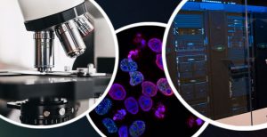
We use ‘tracers’ to find cancer proteins called ‘biomarkers’ in a sample of tissue. These tracers are antibodies that attach to biomarker proteins when added to the sample. We add dyes to the tracers so that we can clearly see them with a microscope. This shows us which biomarkers the tissue has and how much of each is present. We then take digital pictures and use computers to analyze them and create a ‘fingerprint’ of the tissue. These tissue fingerprints tell us if all cells in a tumour have the same or different biomarkers, and how much of each biomarker is present. We can then use computer learning tools we develop to group cancers based on their patterns of biomarkers.
The insights from our studies and the imaging tools we develop are improving how we see and understand cancer. This will help each cancer patient get the right treatment at the right time.
More about Imaging Validation Core
IVC lab at Sunnybrook. The Imaging Validation Core (IVC) is focused on the development and validation of reliable quantitative prognostic and predictive biomarkers and validation of new imaging modalities against ground truth provided by histopathology and molecular profiling. This work is carried out at the Biomarker Imaging Research Lab at the Sunnybrook Research Institute.
Expertise and capabilities: In addition to the biomarker multiplexing capability, the lab offers full range of digital whole-mount histopathology techniques, whole mount immunohistochemistry and development of image analysis tools and digital pathology applications and biomarker multiplexing. The lab offers access to imaging-pathology validation services and resources for clinical trials and pilot projects.
Principal Investigator:
Dr. Martin Yaffe, Senior Scientist, Physical Sciences, Tory Family Chair in Cancer Research, Sunnybrook Research Institute; Chief Scientific Officer, CIMTEC; Professor, Departments of Medical Biophysics and Medical Imaging, University of Toronto
Contact:
Yulia Yerofeyeva
Website:
https://sunnybrook.ca/
What we do?
The Quantitative Imaging for Personalized Cancer Medicine (QIPCM) program supports imaging research at hospitals in Ontario and around the world. We provide tools and services for research groups to support them in conducting imaging-based clinical trials.
Why we do it?
Imaging is a useful tool in cancer research. It can tell us where a person’s tumour is, and also measure things like the size of a tumour, how much blood flow reaches the tumour, or even how much drug is reaching the tumour. Clinical trials that use imaging are building knowledge about cancer and can lead to new and improved treatment options for patients. Our work ensures these trials are of the highest quality and contributes to patients having the latest cancer treatments.
How we do it?
Our team provides many tools and services. Some of these tools and services protect patient privacy. Others collect and save images from many hospitals globally into one space. And still others check the images to make sure they are good quality and can analyze this data. QIPCM supports advanced imaging techniques as well, some of which are only used in research studies. We are always working to build more tools to support imaging techniques. QIPCM’s tools and services ensure that imaging data is accurate, reliable, and consistent.
More about QIPCM
QIPCM provides end-to-end data management and medical imaging solutions to help clinical trials achieve improved consistency and reliability in study data. QIPCM has established the required infrastructure and support services for international multi-centre clinical trials. QIPCM is based out of University Health Network.
Expertise and capabilities: QIPCM services for clinical trials includes a centralized data repository with remote access for multi-centre clinical trials, data anonymization and transfer, scanner quality assurance, imaging protocol development and image analysis.
Principal Investigator
Dr. David Jaffray, Senior Scientist, Princess Margaret Cancer Centre ; Executive VP Technology and Innovation, University Health Network; Director, Research Institute, Techna Institute for the Advancement of Technology for Health
Contact
Julia Publicover